
Orthopantomography, also known as panoramic dental radiography, is a valuable tool in the field of dentistry that aids in the diagnosis and treatment of various dental conditions. This comprehensive guide aims to provide a deeper understanding of orthopantomography, its benefits, limitations, symptoms, causes, and more. In this article, we will delve into the intricacies of this imaging technique, uncover the symptoms and causes associated with it, and explore its significant role in the field of dentistry. Whether you are a dental professional seeking to enhance your knowledge or a patient curious about your dental health, this article will equip you with the essential information needed to better comprehend orthopantomography and its implications in diagnosis and treatment.
1) "Understanding Orthopantomography: A Comprehensive Guide to Diagnosis and Treatment"
Orthopantomography, also known as dental panoramic radiography, is a diagnostic imaging technique used to obtain a comprehensive view of the teeth, jaws, and surrounding structures. It provides valuable information for dentists and oral healthcare professionals to assess the condition of the oral cavity and plan appropriate treatments. In this section, we will delve into the details of orthopantomography, discussing its purpose, procedure, benefits, and how it aids in diagnosis and treatment.
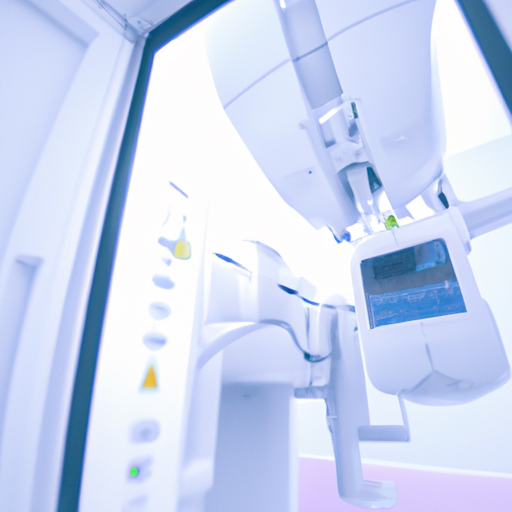
Orthopantomography is a non-invasive and painless procedure that involves taking a single panoramic X-ray image of the entire mouth. It captures a wide view of the upper and lower jaws, including the teeth, supporting structures, temporomandibular joints, and sinuses. The resulting image displays a detailed overview, highlighting any abnormalities or issues that may not be visible in regular dental X-rays.
The primary purpose of orthopantomography is to aid in the diagnosis and treatment planning of various dental conditions. Dentists use the images to identify dental caries (cavities), periodontal disease, impacted teeth, cysts, tumors, and other abnormalities. By visualizing the entire oral cavity, orthopantomography helps dentists evaluate the alignment of teeth, detect bone loss, assess the development of permanent teeth in children, and evaluate the need for orthodontic treatment.
During the procedure, the patient is positioned in front of the imaging device, and a specialized X-ray machine rotates around their head. The machine emits a controlled amount of radiation to capture the panoramic image. To ensure a clear and accurate image, patients are typically required to remove any jewelry, eyeglasses, or metal objects that may interfere with the X-ray.
The benefits of orthopantomography are numerous. Firstly, it provides a comprehensive view of the entire oral cavity, allowing dentists to identify multiple dental issues simultaneously. This saves time and resources, as separate X-rays for different areas are not required. Furthermore, orthopantomography is a valuable tool in treatment planning. Dentists can accurately measure bone
2) "Unveiling the Symptoms and Causes of Orthopantomography: What You Need to Know"
Orthopantomography, also known as panoramic dental radiography, is a diagnostic imaging technique used in dentistry to obtain a comprehensive view of the jaws, teeth, and surrounding structures. This non-invasive procedure is widely used for diagnosing and evaluating various dental conditions. Understanding the symptoms and causes of orthopantomography is crucial for both patients and dental professionals.
Symptoms of orthopantomography are generally related to the specific dental conditions being investigated. Patients may experience symptoms such as toothache, jaw pain, difficulty chewing, gum swelling, or abnormal tooth alignment. These symptoms may prompt a dentist to recommend orthopantomography to obtain a clearer picture of the underlying issue.
One of the primary causes for requiring orthopantomography is to identify dental abnormalities or pathologies. These can include impacted teeth, cysts, tumors, infections, periodontal diseases, or fractures. Orthopantomography allows dentists to visualize these conditions in detail, aiding in accurate diagnosis and treatment planning.
Another common cause for orthopantomography is dental implant planning. Dental implants are artificial tooth roots used to replace missing teeth. Before the implantation process, dentists need to evaluate the available bone structure, the proximity of nerves, and the presence of any anatomical variations. Orthopantomography provides a comprehensive view of the jawbone, enabling dentists to assess suitability for dental implant placement.
Orthopantomography is also useful in orthodontic treatment planning. Before initiating orthodontic treatment, dentists need to assess the alignment of teeth, the presence of impacted teeth, and the overall condition of the jaw. This imaging technique allows orthodontists to develop a customized treatment plan tailored to the patient’s specific needs.
Furthermore, orthopantomography plays a vital role in detecting temporomandibular joint (TMJ) disorders. TMJ disorders can cause various symptoms, including jaw pain, clicking or popping sounds, limited jaw movement, or headaches. Orthopantomography helps dentists identify any abnormalities in the TMJ, facilitating appropriate treatment options.
3) "Exploring the Benefits and Limitations of Orthopantomography in Diagnosis and Treatment"
Orthopantomography, also known as panoramic dental radiography, is a valuable diagnostic tool used in dentistry to obtain a comprehensive view of a patient’s oral and maxillofacial structures. This imaging technique captures a two-dimensional image of the entire mouth, including the teeth, jaws, temporomandibular joints, and surrounding structures. While orthopantomography offers several benefits in diagnosis and treatment planning, it also has its limitations that should be considered.
One of the primary advantages of orthopantomography is its ability to provide a broad overview of the oral cavity in a single image. This panoramic view allows dentists to assess the position and alignment of teeth, evaluate the supporting bone structures, detect infections or cysts, and identify any abnormalities or pathologies. With this comprehensive visual information, dentists can make accurate diagnoses and develop appropriate treatment plans.
Furthermore, orthopantomography is a non-invasive and relatively quick imaging technique. Patients simply need to stand or sit in a specific position while the machine rotates around their head, capturing the image. This process is comfortable for patients and does not require any injections or exposure to potentially harmful radiation doses. The convenience and ease of orthopantomography make it a preferred method for routine dental examinations and screenings.
Orthopantomography also plays a crucial role in treatment planning, particularly in oral surgeries and orthodontic procedures. Dentists can use these panoramic images to evaluate the position of impacted teeth, plan the extraction of wisdom teeth, assess the need for dental implants or dentures, and monitor the progress of ongoing treatments. Additionally, orthopantomograms are beneficial for determining the presence of temporomandibular joint disorders and evaluating their severity, aiding in the development of appropriate treatment strategies.
Despite its numerous advantages, orthopantomography has certain limitations that should be acknowledged. The two-dimensional nature of the images can sometimes limit the accuracy of measurements, making it challenging to assess precise distances or determine the depth of lesions accurately. Additionally, superimposition of structures may occur, potentially masking certain path
